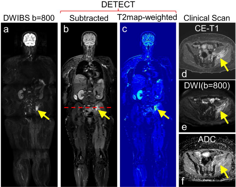
Figure 6.

Whole-body MRI of a 58-year old female patient volunteer with advanced renal cell carcinoma and underwent radiation treatment to the left iliac bone metastatic lesion. DWIBS image at b=800 s/mm2 (a), subtracted DETECT image (b) and the effective T2map-weighted image (c) show conspicuous lesion. Clinical contrast-enhanced fat saturated T1-weighted image of the same patient reveals an enhancing left iliac bone lesion (d, yellow arrow), which also appeared hyperintense on clinical DWI image with b = 800 s/mm2 (e, yellow arrow), and ADC map (f) (calculated from 4 b-values; 0, 50, 400, 800 s/mm2), indicative of residual tumor with post-radiation effects.
