
Figure 1. Mitochondrial content with CCA.
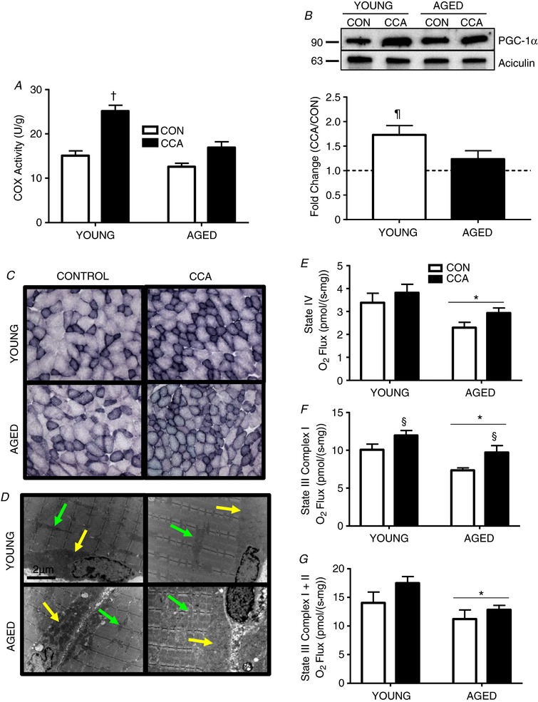
A, COX activity in vehicle‐treated young and aged muscle following CCA (n = 6–8). B, PGC‐1α protein expression expressed as fold change of CCA over CON (n = 5–6). C, SDH staining was performed on 10 μm sections. A representative image based on the assessment of two animals per condition is shown at 10× magnification. D, representative electron micrographs of mitochondria based on the assessment of two animals per condition (young and aged VEH‐treated muscle) are shown. Yellow arrows denote SS mitochondria and green arrows indicate IMF mitochondria. E–G, respiration was measured on intact saponin‐ permeabilized muscle fibre bundles for basal (E) and maximal (F and G), respiration states (n = 5–8). Data are presented as means ± SEM. † P < 0.05 CCA vs. all other conditions; ¶ P < 0.05 CCA vs. CON; * P < 0.05 main effect of age; § P < 0.05 main effect of CCA. CON, control; CCA, chronic contractile activity, COX; cytochrome oxidase; SDH, succinate dehydrogenase.