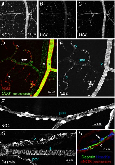
Figure 1. Morphological characteristics of mural cells in the microvasculature of bladder suburothelium.

In a non‐fixed mucosal preparation of NG2‐DsRed mouse bladder, the DsRed fluorescence signal revealed the NG2‐expressing mural cells in the microvascular network (A). A single plane image of the same area showed an extensive network of capillary pericytes just beneath the urothelium (B), whereas another single plane image visualized arteriolar tree branching capillaries in the outermost part of the lamina propria (C). In another lamina propria preparation processed for immunohistochemistry against the endothelial marker CD31, arteriolar smooth muscle cells expressed a strong NG2 signal (a), whereas no fluorescence was detected in venular pericytes (v) (D and E). Capillary pericytes (c) also expressed a strong NG2 signal, whereas pericytes in the postcapillary venule (pcv) showed faint NG2 signals (D and E). In a different lamina propria preparation, pericytes in the precapillary arteriole (pca) had an oval cell body expressing a strong NG2 signal and extended circumferentially‐arranged processes with weaker NG2 signals (F). The stellate shaped morphology of pericytes in a venule (v) and a postcapillary venule (pcv) were revealed by desmin immunohistochemistry, whereas circumferentially‐arranged processes were evident in the arteriole (a) (G). A capillary pericyte and endothelium (c) were visualized by their immunoreactivity for desmin (green) and eNOS (red), respectively (H). Nuclear staining with Hoechst (blue) indicated the pericyte cell body (arrow) extending longitudinally‐oriented bipolar long processes. The scale bar in (A) also refers to (B) and (C), and that in (D) also refers to (E). [Color figure can be viewed at http://wileyonlinelibrary.com]
