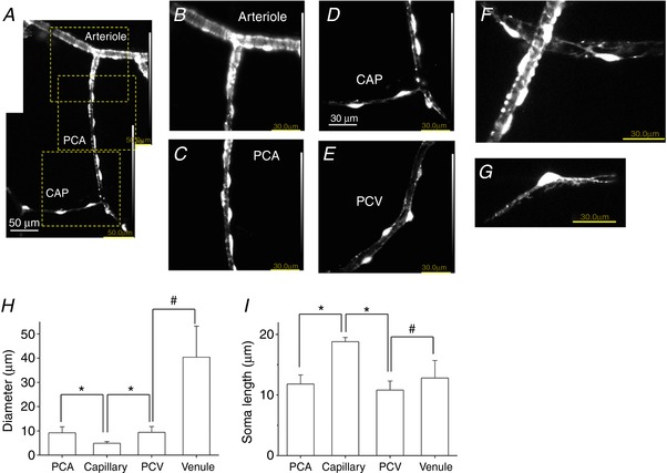
Figure 2. Morphological characteristics of mural cells in the microvasculature network for Ca2+ imaging.

In a lamina propria preparation of NG2‐DsRed mouse bladder used for Ca2+ imaging, an arteriole covered by circumferentially‐arranged NG2(+) SMCs branched into a PCA where NG2(+) pericytes lined along the boundaries of the PCA wall (A). The PCA continued to a capillary (CAP) where NG2(+) pericytes were more sparsely distributed (A). An expanded image of the top dotted square in (A) revealed circumferentially‐arranged NG2(+) arteriolar SMCs (B). An expanded image of the middle dotted square in (A) showed NG2(+) PCA pericytes with an oval shaped cell body extending circumferentially‐oriented processes (C). An expanded image of the bottom dotted square in (A) showed NG2(+) CAP pericytes with an oval shaped cell body extending bipolar longitudinal processes (D). Pericytes in PCV had a short oval cell body that expressed a faint NG2 fluorescence (E). Note that luminance was increased for clarity. In another lamina propria preparation, NG2(+) pericytes extended circumferentially‐oriented processes wrapped around PCA, whereas stellate‐shaped NG2(+) pericytes were distributed in PCV (F). A capillary pericyte expressed strong NG2 fluorescence in the cell body and extended NG2(+) bipolar processes (G). Microvascular diameter (H) and soma length of pericytes (I) in different microvascular segments are summarized. Data are the mean ± SD. NS, not significantly different. *Significantly different from the values of capillary (unpaired Student's t test, P < 0.05). #Significantly different between the values of PCV and venule (unpaired Student's t test, P < 0.05). [Color figure can be viewed at http://wileyonlinelibrary.com]
