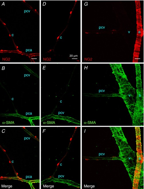
Figure 4. Immunoreactivity for α‐SMA in different segments of suburothelial microvessels of NG2 DsRed mouse bladder.

Pericytes in a PCA expressed a red NG2 signal (pca), as well as α‐SMA immunoreactivity (green), whereas the NG2‐expressing pericytes in a capillary (c) branching from the PCA were α‐SMA‐negative (A–C). In the same micrographs, NG2(+) pericytes in a postcapillary venule (pcv) expressed faint α‐SMA immunoreactivity. NG2(+) pericytes in a postcapillary venule (pcv) but not those in a connecting capillary (c) exhibited α‐SMA immunoreactivity (D–F). Pericytes in a larger venule without NG2 signal (v) showed strong α‐SMA‐immunoreactivity, and a connecting postcapillary venule with faint NG2 signal (pcv) exhibited weaker α‐SMA‐immunoreactivity (G–I). Smooth muscle cells in an arteriole (a) expressing a bright red NG2 signal showed strong α‐SMA immunoreactivity. The scale bar in (A) = 20 μm also refers to (B) to (I). [Color figure can be viewed at http://wileyonlinelibrary.com]
