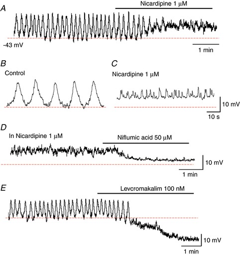
Figure 9. Electrical properties of venular pericytes.

In a venule where pericytes periodically generated slow waves, nicardipine (1 μm) depolarized the membrane by ∼10 mV and prevented the slow wave generation leaving STDs (A). The traces with an expanded time scale showed slow waves in control (B) and STDs in nicardipine (C). In the same venule, subsequent niflumic acid (1 μm) hyperpolarized the membrane by ∼10 mV and abolished STDs (D). In another venule where slow waves were generated, levcromakalim (100 nm) hyperpolarized the membrane by ∼15 mV and abolished slow waves (E). [Color figure can be viewed at http://wileyonlinelibrary.com]
