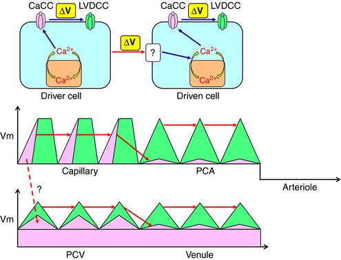
Figure 11. Proposed model of voltage‐dependent coupling cytosolic Ca2+ oscillators.

Driver cells are capable of operating a ‘spontaneous’ cytosolic Ca2+ oscillator that is linked with CaCCs to cause a depolarization triggering the activation of LVDCCs. By contrast, driven cells require depolarizing input from driver cells. The depolarizing input activates cytosolic Ca2+ oscillator presumably by voltage‐dependent IP3 production resulting in the activation of CaCCs and VDCCs. Capillary pericytes act as ‘driver cells’ and generate CaCC‐depolarizations that are sufficiently large to couple with each other. PCA pericytes are driven by a spread of the depolarization via gap junctions that activate LVDCCs to couple with each other. The hyperpolarized membrane of arteriolar SMCs prevents the sequential spread of oscillations. Capillary pericytes may also electrically drive PCV pericytes. Upon the background CaCC conductance, a population of PCV pericytes could act as driver cells and generate CaCC‐depolarizations, although they require LVDCC activation to couple with each other. Venular pericytes could be driven by a spread of the depolarization via gap junctions that activate LVDCCs to couple with each other. [Color figure can be viewed at http://wileyonlinelibrary.com]
