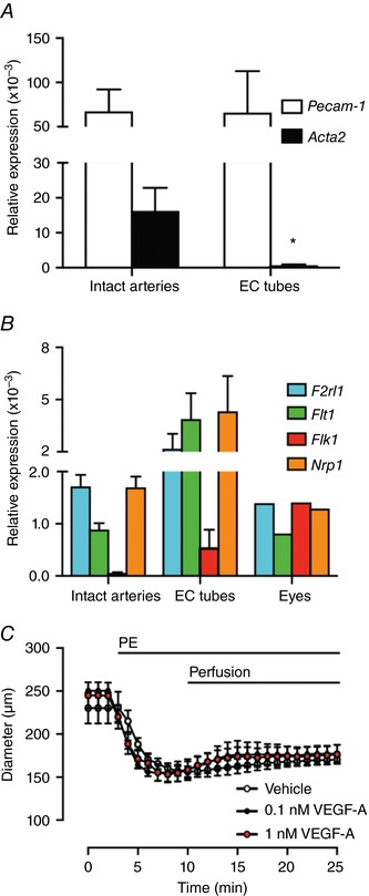
Figure 1. VEGF‐A receptor expression in mouse mesenteric resistance arteries.

A, Pecam‐1 and Acta2 expression in intact arteries and isolated EC tubes. B, Flt1, Flk1 and Nrp1 expression (n = 4 animals). Eyes from a single animal were used as a positive control for the VEGF‐A receptors. C, arteries were pre‐constricted with PE. When pumped into the lumen of arteries, neither vehicle, nor VEGF‐A dilated the artery within 15 min (n = 3–4). * P < 0.05 compared to intact arteries.
