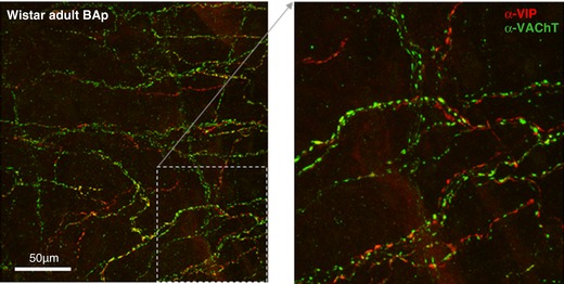
Figure 8. Juxtapositioning of cholinergic and peptidergic autonomic fibres innervating the vertebrobasilar arteries.

Peptidergic and cholinergic markers sometimes, but far from always, co‐localise in the same fibres. Though the two fibre types tend to run in parallel, in the majority of fibres there is a clear separation of the two markers. Confocal image of α‐VIP‐594 and α‐VAChT‐488 immunofluorescence in the posterior basilar artery in an adult Wistar rat. Flattened confocal 100 μm z‐stack and an excerpt shown at higher power. [Color figure can be viewed at http://wileyonlinelibrary.com]
