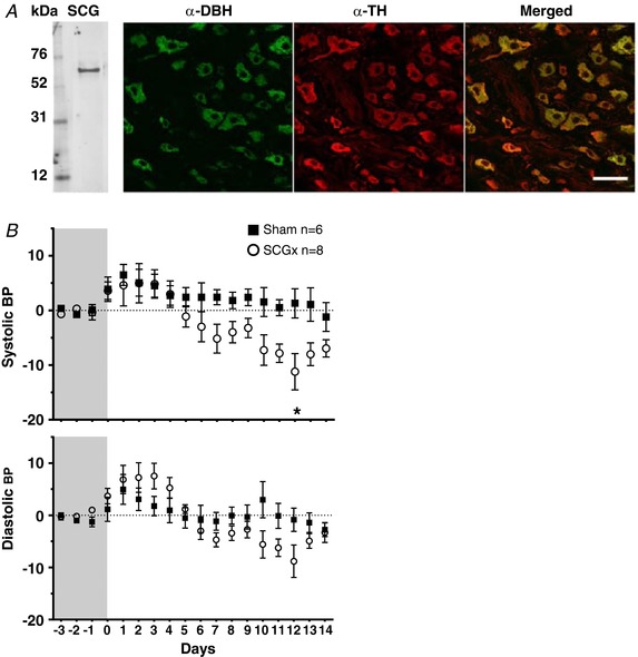
Figure 13. Identification of excised tissues as superior cervical ganglion and development in blood pressure following excision.

A, the excised tissue was positive for tyrosine hydroxylase (TH) protein as shown by western blotting. Co‐localisation study of α‐DBH and α‐TH showed that cell bodies within the tissue are positive for both tyrosine hydroxylase as well as dopamine‐β‐hydroxylase. All DBH fluorescence is localised to TH fluorescence suggesting that they mark the same structures. Scale bar: 50 μm. B, the blood pressure measurements (in mmHg) were obtained using DSI telemetry. After recording 3 days of baseline, animals underwent either sham or SCGx treatment and blood pressure was recorded continually for 14 days. The traces are presented as difference from the baseline. The systolic and diastolic blood pressure traces of ganglionectomised and sham operated animals are presented as changes from baseline. [Color figure can be viewed at http://wileyonlinelibrary.com]
