Figure 7.
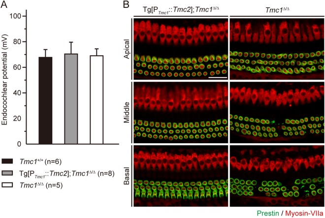
Secondary effects of Tg[PTmc1::Tmc2] in Tmc1∆/∆ mice. (A) Normoxic endocochlear potentials (mean ± SD) of Tg[PTmc1::Tmc2]; Tmc1∆/∆ (gray bar), Tmc1∆/∆ (white bar) and wild-type (Tmc1+/+, black bar) mice at P16. Normoxic endocochlear potentials of both Tg[PTmc1::Tmc2]; Tmc1∆/∆ and Tmc1∆/∆ mice were not significantly different from those of wild-type mice (one-way ANOVA, P > 0.05). (B) Prestin expression in Tg[PTmc1::Tmc2]; Tmc1∆/∆ (n = 4 mice) and Tmc1∆/∆ cochleae (n = 4 mice) at P16. Prestin immunoreactivity38 was identified along the perimeter of outer HCs from the apical to basal turns of both Tg[PTmc1::Tmc2]; Tmc1∆/∆ and Tmc1∆/∆ cochleae at P16. In Tmc1∆/∆ cochleae, outer HCs were degenerating in the middle to basal turns. Inner and outer HCs were counter stained with myosin-VIIa (red; scale bars, 25 µm).

