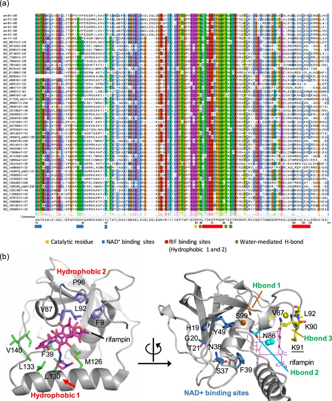
Figure 1.
Integrated analysis of rifamycin ADP-ribosyltransferase (Arr). (a) The sequence conservation of rifamycin ADP-ribosyltransferase (Arr). The residues that are conserved more than 78% of the sequences are highlighted based on the clustalw color scheme. The genes selected for experimental validation are shown as wd1-wd4. (b) Proposed key residues and their molecular interactions in the rifampin-binding sites of Arr-ms structure (PDB 2HW2). In Arr-ms, the variable X residue in M[R|K][D|E]XL motif is Gly129, and its position is highlighted with a red arrow. Water molecules are depicted with spheres.

