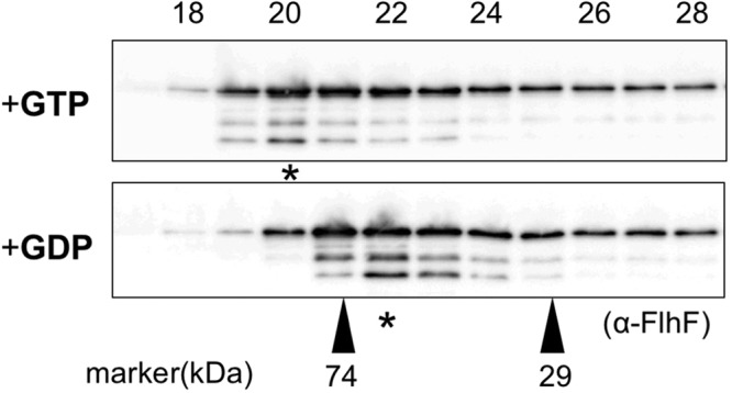
Figure 6.

FlhF forms a dimer in the presence of GTP. FlhF was incubated in the presence of either 1 mM GTP or GDP and analyzed by gel filtration chromatography using buffer containing either 1 mM GTP or GDP. The upper panel shows the profile in the presence of GTP, and the lower panel shows it in the presence of GDP. Immunoblotting was performed for each fraction using the anti-FlhF antibody. The regions of interest were cropped from the immunoblotting. The peak fraction containing FlhF is indicated by the asterisk. The molecular size markers were carbonic anhydrase (29 kDa) and conalbumin (74 kDa).
