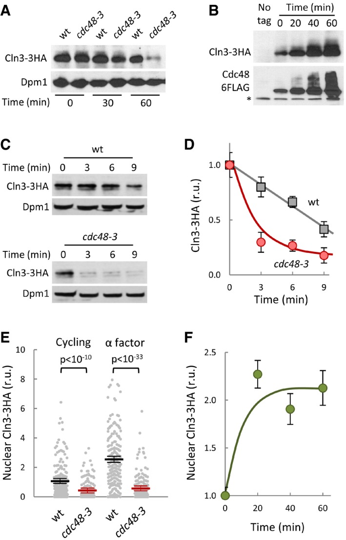
Figure 2. Cdc48 prevents degradation and stimulates nuclear accumulation of Cln3.

- Cln3‐3HA levels in wild‐type and cdc48‐3 cells at the indicated times after transferring cells to the restrictive temperature (37°). Dpm1 is shown as loading control.
- Cln3‐3HA levels at the indicated times after addition of β‐estradiol to induce the GAL1 promoter in GAL1p‐CDC48‐6FLAG cells expressing the Gal4‐hER‐VP16 (GEV) transactivator. The bottom panel shows Cdc48‐FLAG levels and a cross‐reacting band (*) that serves as loading control.
- Cln3‐3HA stability in Cdc48‐deficient cells. After being transferred to the restrictive temperature (37°C) for 30 min, cycloheximide was added to wild‐type and cdc48‐3 cells, which were collected at the indicated times to determine Cln3‐3HA levels. Dpm1 is shown as a loading control.
- Quantification of Cln3‐3HA levels in wild‐type (squares) and cdc48‐3 (circles) cells from immunoblot analysis as in (C). Mean values (N = 3) and confidence limits (α = 0.05) for the mean are shown.
- Nuclear accumulation of Cln3‐3HA in individual wild‐type or cdc48‐3 cells either cycling or arrested in late G1 with α factor as measured by semiautomated quantification of immunofluorescence levels in both nuclear and cytoplasmic compartments after 60 min at the restrictive temperature. Values were made relative to the average value obtained from wild‐type cells. Mean values (N = 200, thick horizontal lines), confidence limits (α = 0.05, thin lines) for the mean and P‐values obtained from t‐tests are also shown.
- Nuclear accumulation of Cln3‐3HA at the indicated times after overexpressing Cdc48 as in (B). Immunofluorescence analysis and calculations of relative mean values (N = 200) and confidence limits (α = 0.05) for the mean are as in (E).
Source data are available online for this figure.
