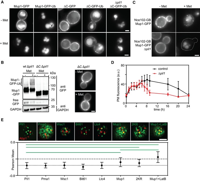
Influence of the in‐frame fusion of an ubiquitin (Ub) moiety to Mup1 and ΔC on PM localization and turnover. V = vacuole.
Western blot showing Mup1 and ΔC ubiquitination in Δ
pil1. Samples were prepared 5 min after addition of Met. Loading control as in Fig
1C. Representative images of indicated conditions are shown to illustrate defect in endocytosis of ΔC.
Artificial anchoring of Mup1 to Nce102 in the presence or absence (Δpil1) of MCC and Met, respectively.
Mup1 internalization during long‐term growth in the absence of Met.
Degree of colocalization of endocytic events (shown by the late endocytic marker Abp1‐RFP) with various PM domains (GFP fusions). Representative two‐color TIRFM images of the indicated protein pairs are shown. Pil1 represents eisosomes, Pma1 the MCP, Wsc1 the MCW (membrane compartment occupied by Wsc1), Bit61 the MCT, and Ltc4 the MCL (membrane compartment of Ltc3/4). Cells were grown in Sc‐Met and imaging performed after addition of 100 μM Met. LatB was used at a concentration of 100 μM to stabilize endocytic sites. The dotted line is used as a guide to the eye.
Data information: Values represent means ± SD,
n = 50–150 (D)
n = 25–80 (E). Green lines indicate significantly different data sets. An overview of the performed statistics can be found in
Table EV3. Scale bars: 2 μm. All measured values are listed in
Table EV1.
Source data are available online for this figure.

