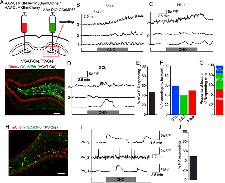
Figure 3. MC commissural projections excite a subset of dentate GABA interneurons.

(A) Top: Viral injection scheme in VGAT-Cre (or PV-Cre) mice. Bottom: commissural projections and GCaMP6f expression in the contralateral DG of a VGAT-Cre mouse. Scale bar 100 μm.
(B) (B-D) Representative ΔF/F signals in contralateral VGAT+ interneurons from (B) subgranular zone (SGZ), (C) hilus, or (D) granule cell layer (GCL) upon CNO application.
(E) Percent of GCaMP6f+ GABA interneurons that increased Ca2+ events (⩾ 50%) upon chemogenetic activation of MC commissural projections.
(F) Percent of responsive VGAT+ cells residing within SGZ, hilus, or GCL.
(G) Distribution of the responders among the SGZ, hilus, and GCL.
(H) Commissural projections and GCaMP6f expression in the contralateral DG of a PV-Cre mouse. Scale bar 100 μm.
(I) Representative ΔF/F signals in contralateral PV+ interneurons upon CNO application.
(J) Percent of GCaMP6f+ PV interneurons that increased Ca2+ events (⩾ 50%) in response to chemogenetic activation of MC commissural projections.
