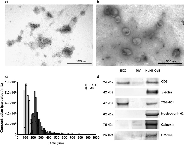
Fig. 1.
Characterisation of urinary exosomes isolated by ultracentrifugation. Transmission electron microscopy (TEM) micrographs of extracellular vesicle isolations stained with uranyl acetate in a exosomes and b microvesicles, bar represents 500 μm. c The size distribution of urinary exosomes and microvesicles by tunable resistive pulse sensing analysis of 500 events per sample (n = 4). d Western immunoblotting of exosomes and microvesicles isolated from urine using exosomal markers, including Tsg101 and CD9, and non-exosomal markers including calnexin, nucleoporin, p62, GM-130 and β-actin. Whole-cell lysates from HuH7 cells were loaded as a positive control

