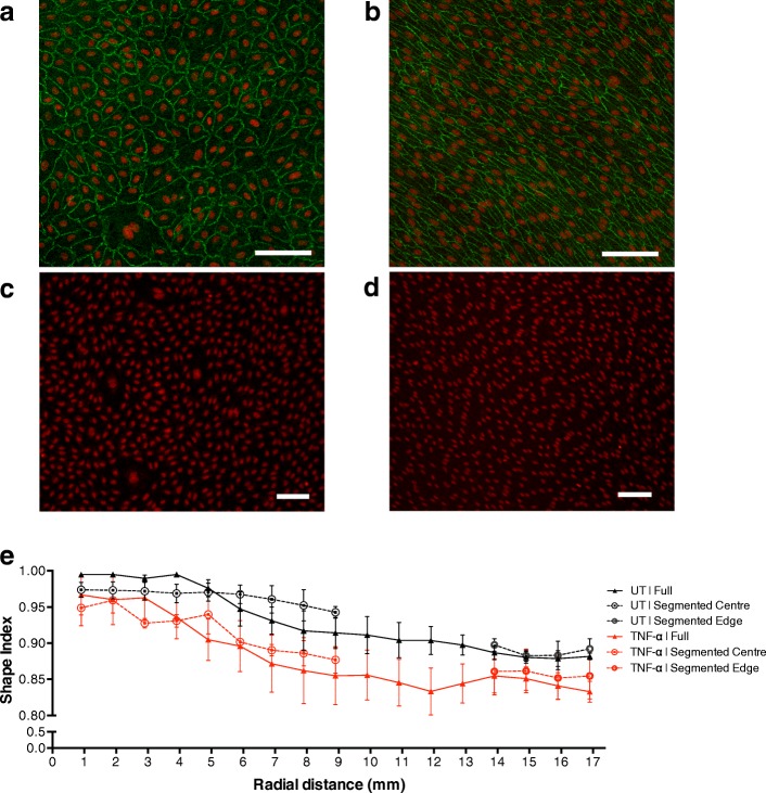
Fig. 4.
Nuclear (red) stain shows the morphology of sheared HUVEC (a) in the centre and (b) at the edge of a full well, and (c) in the centre and (d) at the edge of a segmented well (scale bar = 100 μm). a and b also show cell outlines, delineated by immunostaining of ZO-1. Note the alignment and elongation of cells at the edge but not at the centre, and the lack of difference between full and segmented wells. e No significant difference in nuclear Shape Index, indicating roundness, between HUVEC grown in full wells and segmented wells was seen for untreated or TNF-α treated HUVEC. Cells were more elongated near the edge of the well. A tendency for greater elongation in TNF-α-treated HUVEC was not consistently significant across locations

