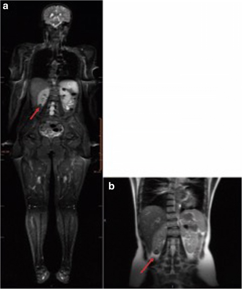
Fig. 1.

Detection of a renal lesion by WB-MRI in LFS patient, Y0102T000. a STIR image of a 19-year-old with cortical oval kidney lesions in the right lower pole (16 mm) and in the left upper pole (20 mm) (indicated with arrows). b An abdominal MRI detected a complex nodule in the lower third of the right kidney, and it measured 23 × 18 × 18 mm3 (indicated with an arrow)
