
Fig. 3.
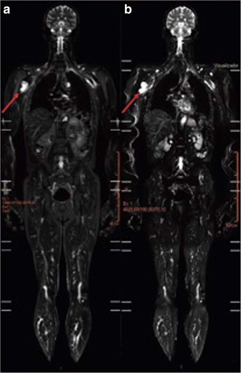
WB-MRI of LFS patient, Y0012T012. A STIR image (a) and diffusion-weighted imaging (b) from WB-MRI. An expansive lesion (48 × 34 mm2) in contact with the short head of the biceps was detected in the proximal segment of the right humerus. The lesion exhibited a hyperintense signal on STIR and restriction on diffusion-weighted imaging scans (indicated with arrows)