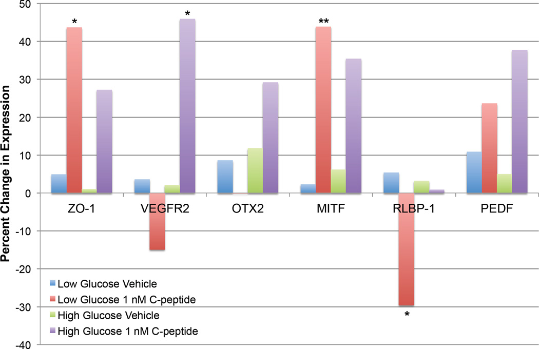
Fig. 6.
C-peptide and retinal pigment epithelium (RPE) cell gene expression changes in a transdifferentiation assay. ARPE-19 cells were incubated in either low (5.5 mM) or high (25 mM) glucose for 30 days with or without 1 nM C-peptide. Medium +/− C-peptide was replenished weekly. To evaluate C-peptide-induced changes in gene expression, cells were lysed, RNA isolated, and changes in gene expression evaluated by quantitative PCR using primers specific against genes produced by RPE cells, including the tight junction protein zona occludens 1 (ZO-1), the VEGF receptor 2 (VEGFR2), the transcription factor orthodenticle homeobox 2 (OTX2), the transcription factor microphthalmia-associated transcription factor (MITF), which is involved in regulating RPE cell identity, retinaldehyde-binding protein 1 (RLBP-1), and pigment epithelium-derived factor (PEDF). Under low glucose conditions, C-peptide significantly altered the mRNA expression of several genes, including ZO-1, MITF, and RLBP-1. However, this effect was lost in cells cultured in high glucose, suggesting that C-peptide-induced changes in gene expression are dependent upon glucose concentration, perhaps reflecting an interaction with AMPK-mediated signaling cascades. *P ≤ 0.05, **P ≤ 0.01 versus vehicle-treated control.

