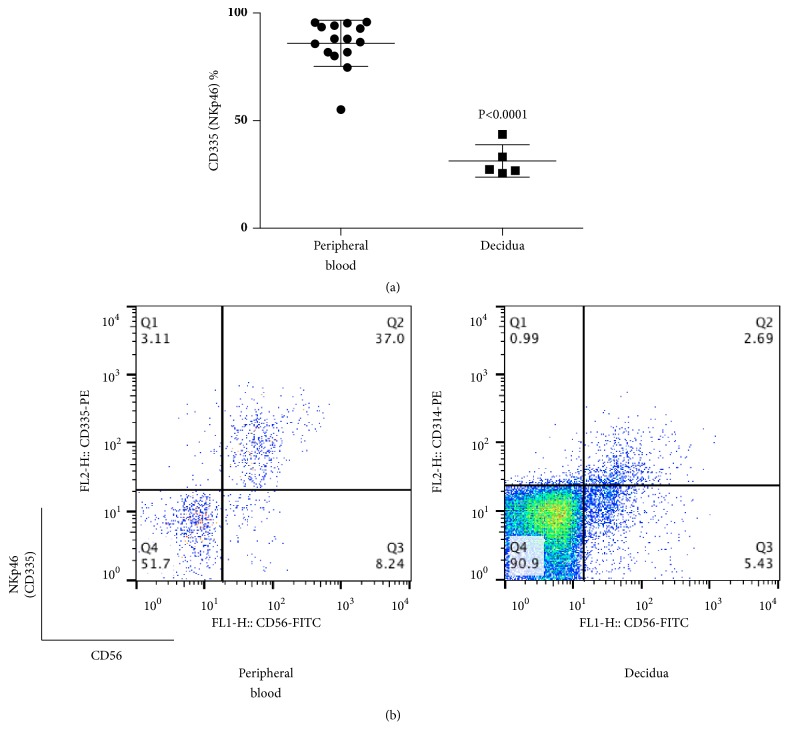
Figure 4.
(a) Percentage of CD335 (NKp46)+ cells among CD56+CD3− cells at term pregnancy in peripheral blood and decidua. The percentage of CD335 (NKp46)+ lymphocytes in decidua was lower than in peripheral blood. (b) Representative dot plot analysis of CD335 (NKp46)+ cells among CD56+CD3- lymphocytes taken from term pregnancy by flow cytometry. The gate was set around the CD3- lymphocytes to analyze the CD335+CD56+ NK cells.

