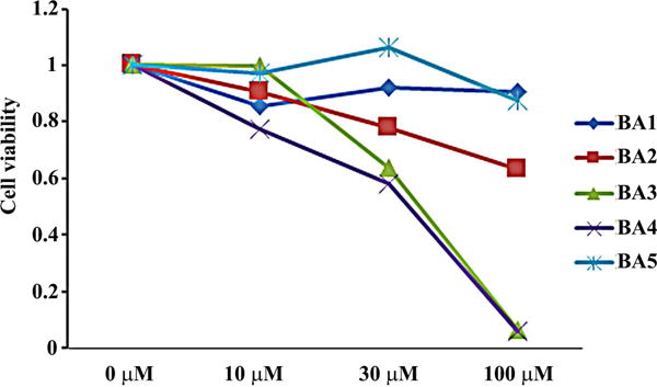
Figure 3.

Cytotoxicity of betulinic acid and its ionic derivatives in 43D cells. (A) 43D cells were treated with different doses of betulinic acid and its ionic derivatives for 48 hrs. The effect of these compounds on cell growth was measured by the AlmarBlue cell proliferation assay. The figure illustrates the relative cell proliferation to the control. The averages of two experiments are shown.
