Figure 1.
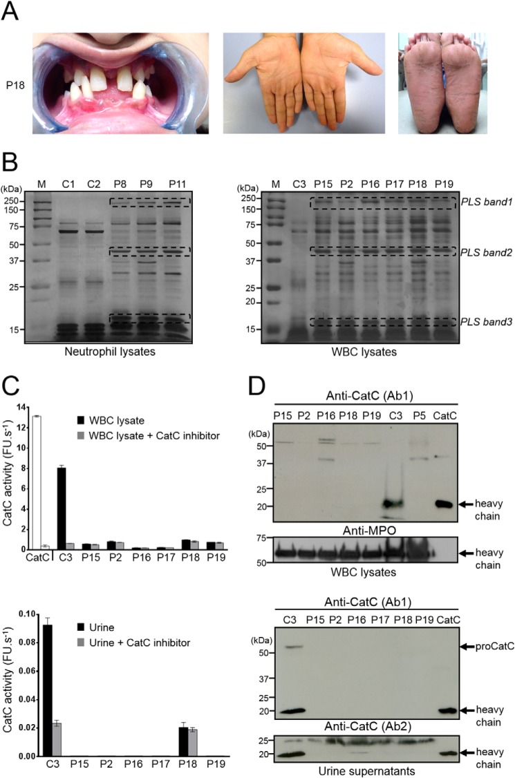
CatC in biological samples of PLS patients. A, characteristic dental and palmoplantar features of PLS (patient 18, P18). Photos show early loss of teeth and hyperkeratosis of the palms and soles. B, neutrophil and WBC lysates from PLS patients and from healthy controls, lysed in 50 mm HEPES buffer, 750 mm NaCl, 0.05% Nonidet P-40, pH 7.4, during 5 min at room temperature and analyzed by SDS-PAGE (12%)/silver staining under reducing conditions (10 μg/lane) strongly differ by their protein profile. Lane M shows protein molecular weight size markers. PLS bands 1, 2, and 3 correspond to proteins that are not cleaved by NSPs in PLS WBC lysates but are hydrolyzed in WBC lysates from healthy controls. Similar results were found in three independent experiments. C, measurement of CatC activity in WBC lysates (10 μg of protein) (top) and concentrated urines (bottom) in the presence or not of a selective CatC inhibitor. The residual proteolytic activity was not inhibited by the CatC inhibitor, which demonstrates the absence of CatC activity in PLS samples. Bars, mean ± S.D. (error bars) of experiments performed in triplicates. D, Western blot analysis of WBC lysates (10 μg of protein) and concentrated urines of PLS samples and controls using anti-CatC antibodies shows the absence of the CatC heavy chain in all PLS samples. The urines were collected and analyzed as in Hamon et al. (34). Similar results were found in three independent experiments. C, control; P, PLS patient; FU, fluorescence units.

