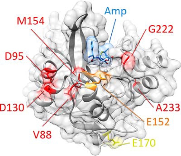
Figure 2.

Position of residues mutated in clinically isolated variants of NDM. NDM is shown as a ribbon diagram with zinc atoms as spheres, coated by a transparent surface (all in gray). Hydrolyzed ampicillin is shown as sticks, with a transparent surface (blue). Positions mutated in clinically isolated variants are color-coded by the highest resistance variant in which each occurs (low in yellow, medium in orange, and high in red, see Fig. 1). Figure was prepared using Protein Data Bank code 4HL2 for di-zinc NDM-1 bound to hydrolyzed ampicillin.
