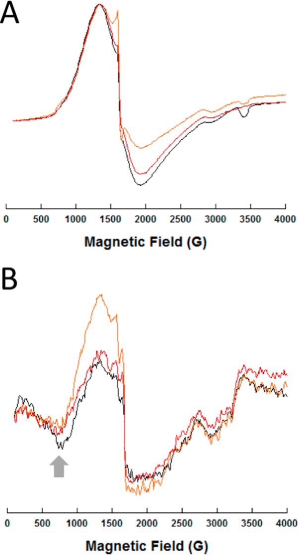
Figure 9.

EPR spectra of representative di-cobalt(II) NDM variants. Spectra for all the NDM variants are given in supporting information. Perpendicular (A) and parallel (B) CW-EPR spectra of the metalloforms (0.5 mm), as labeled, are shown. Sharp spikes at 1600 G are due to a minor contamination from iron. The arrow in the right panel (800 G) indicates the feature associated with cobalt(II)–cobalt(II) coupling. NDM-1 is shown in black, NDM-4 in orange, and NDM-15 in red.
