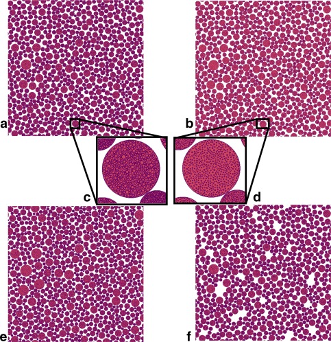
Figure 2.

Cross sections of substrates used in different microstructural scenarios. Space outside cylinders is colored white, space inside cylinders is red, and cell membranes are purple. (a) Baseline substrate. Outer cylinders have radii drawn from a gamma distribution fitted to histology of healthy, wild‐type mouse forelimb tissue. Each cylinder on the substrate contains regularly packed internal cylinders (c). (b) Effect of atrophy scenario 1, in which all internal cylinders are reduced in radius by a fixed factor. (d) The same cylinder as (c) with internal cylinders reduced in radius. Positions are unchanged. (e) A substrate with cylinder radii drawn from a different distribution, fitted to histology of an Mdx mouse model of DMD. Close inspection reveals that these cylinders have a wider range of radii than those in (a), but the packing fractions achieved are similar. Internal structure in (e) is similar to that in (a). (f) Atrophy scenario 2. Here, complete nested cylinders are removed from the baseline arrangement at random. In this example, 10% have been removed. The permeability scenario is not shown, as it is visually identical to the baseline.
