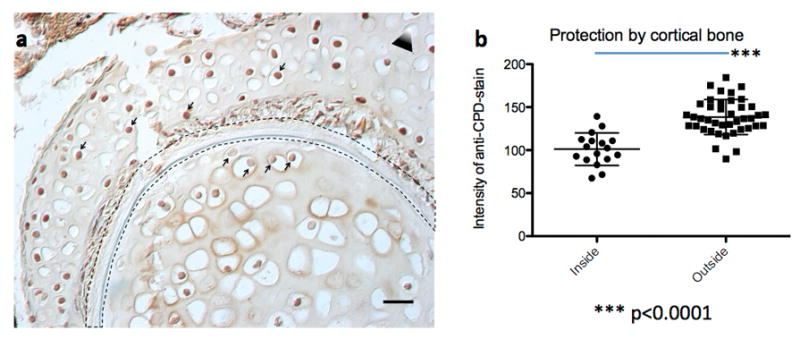
Extended Data Figure 6. Cortical bone protects from UV induced DNA-damage.

(a) Paraffin section of a Dendrobates tinctorius hind leg (from Extended Data Fig. 5a, specimen e) after irradiation with UVB post mortem; the leg was severed from the body and irradiated with UVB. The black dashed outlined represents the cortical bone. Notice the higher staining intensity of the anti-CPD antibody in nuclei within the muscle tissue compared to nuclei within the bone marrow. This part of the leg is not yet hematopoietic (compare to Extended Data Fig. 5e, which shows the hematopoietic marrow in the other leg) but contains chondrocytes. Notice that even the chondrocyte nuclei closest to the cortical bone are stained much less than the cells outside the cortical bone (arrows from below and from above, respectively). The triangle represents the direction of the UV source; white tip towards UV source. The scale bar represents 50 μm. (b) Quantification of grey scale values of nuclei inside (n=17) and outside (n=41) the cortical bone; each data point represents the mean grey value of a 16×16 pixel circle inside the nucleus, the difference is highly significant (unpaired two-tailed t-test, p<0.0001); data are mean with s.d..
