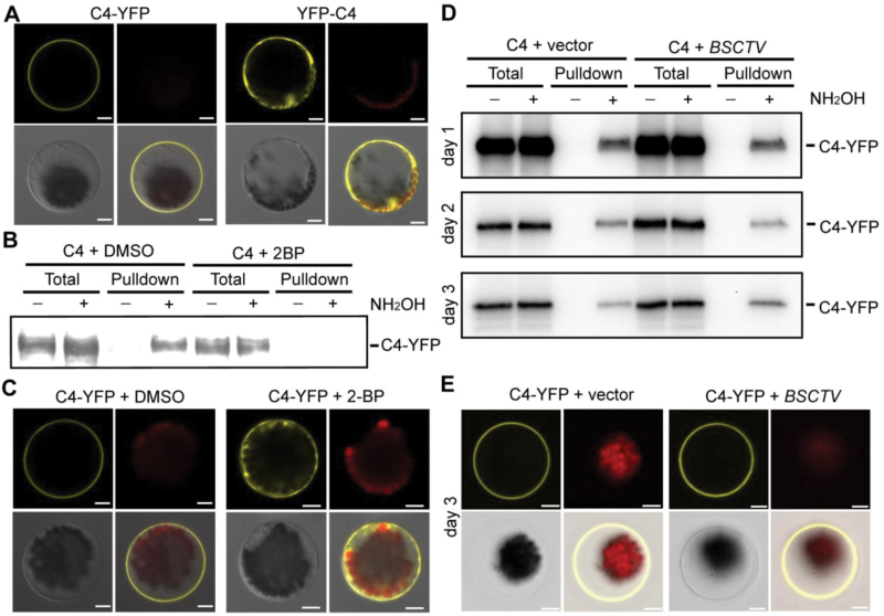
Fig. 1.
S-acylation of BSCTV C4 in plant cells is essential for its membrane localization. (A) C4-YFP-FLAG3His6 or YFP-C4 were expressed in protoplasts and fluorescence was detected by confocal microscopy 18 h after transformation. The YFP signal (yellow), chloroplast autofluorescence (red), bright field (gray), and merged images are shown. Scale bars are 10 µm. (B) Detection of C4 S-acylation in plant cells. C4-YFP-FLAG3His6 was expressed in protoplasts (treated overnight with DMSO or 20 µM S-acylation inhibitor 2-BP) for acyl-resin-assisted capture assays. The signals from the total lysates are shown as the input control, and pulldown indicates the S-acylation proteins captured on the Thiopropyl Sepharose. S-acylation pulldown is dependent on NH2OH. The signals were detected using immunological blotting with an anti-FLAG antibody. (C) C4-YFP-FLAG3His6 was expressed in protoplasts with or without a treatment with 20 µM 2-BP. The fluorescence signals (yellow) were detected using confocal microscopy. Scale bars are 10 µm. (D) S-acylation of C4-YFP-FLAG3His6 was detected in protoplasts without (vector) or with BSCTV infection. The levels of S-acylation were measured 1, 2, and 3 d after infection. (E) Representative images of localization of C4-YFP-FLAG3His6 3 d after BSCTV infection. Scale bars are 10 µm. All results in this figure are representative of three independent experiments.

