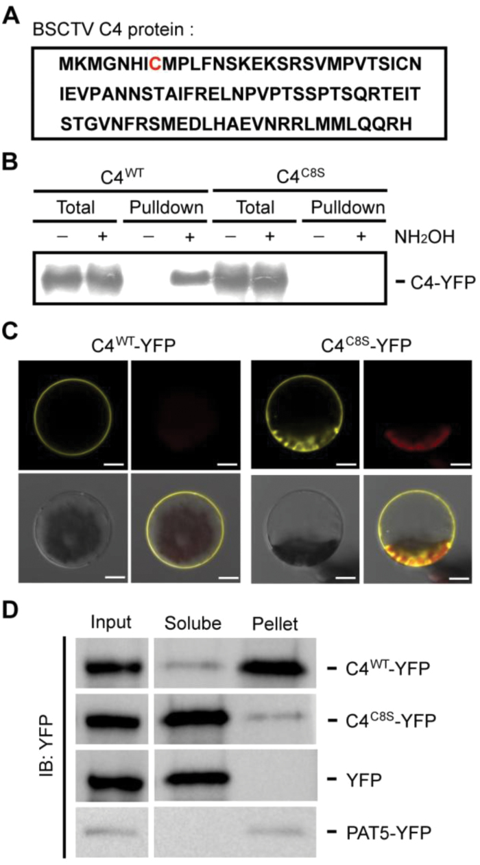
Fig. 2.
Identification of the S-acylation site in BSCTV C4. (A) The predicted S-acylation site in BSCTV C4. The amino acid sequence of C4 is shown and the potential S-acylation site (C8) is highlighted in red. (B) Characterization of the S-acylation site on the C4 protein using acyl-resin-assisted capture assays. The wild-type (WT) or C8S (cysteine to serine) mutant version of C4-YFP-FLAG3His6 was expressed in protoplasts. The total lysate is shown as the input control, and pulldown indicates the S-acylation proteins captured on Thiopropyl Sepharose. The signals were detected using immunological blotting with an anti-FLAG antibody. (C) The localization of the WT or C8S version of C4-YFP-FLAG3His6, detected using confocal microscopy. The YFP signal (yellow), chloroplast autofluorescence (red), bright field (gray), and merged images are shown. Scale bars are 10 µm. (D) Protoplasts expressing the WT or C8S version of C4-YFP-FLAG3His6 were fractionated into soluble and pellet fractions. YFP and PAT5-YFP (a transmembrane protein) were used as controls. The signals were measured from an immunological blot using an anti-GFP antibody. All results in this figure are representative of three independent experiments.

