Fig. 3.
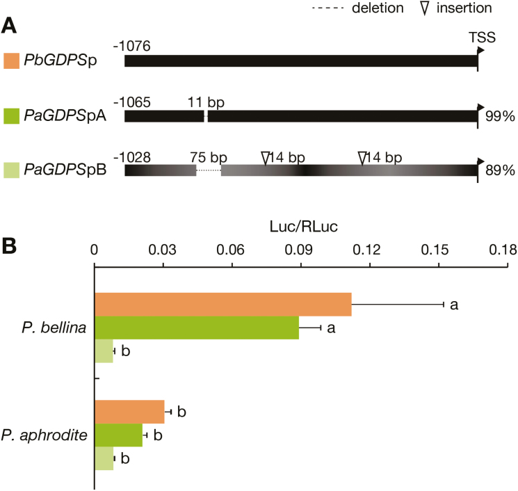
GDPS promoter activities in planta. (A) Sequence differences of three GDPS promoter fragments isolated from P. bellina (PbGDPSp) and P. aphrodite (PaGDPSpA and PaGDPSpB). The numbers indicate base-pairs upstream of the translational start site (TSS). Dotted lines and triangles indicate large-fragment deletions and insertions, respectively, in the two promoter fragments of PaGDPS as compared to PbGDPSp. Comparative similarities are indicated on the right. The numerous substitutions of PaGDPSpB are indicated by the varying shading. (B) Comparative activities of three promoter fragments in the floral tissues of the two Phalaenopsis species as determined by dual-luciferase assays. The activation level is given by the ratio of Luc/RLuc. Data are means (±SE) of three biological replicates. Pairwise comparisons between groups were performed using Tukey’s honestly significant difference test, and different letters indicate significant differences at α=0.05.

