Figure 3.
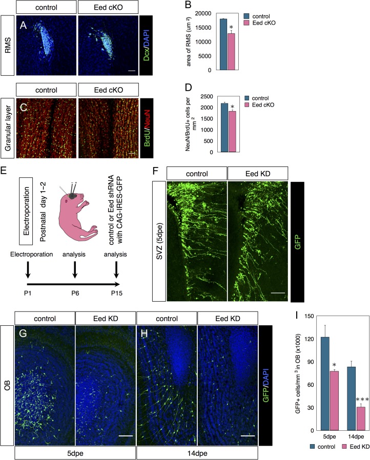
Eed is required for postnatal OB neurogenesis. (A–D) Eed cKO schedule as in Fig. 2F. (A) Immunostaining for Dcx in the RMS. (B) Quantification of surface area occupied by Dcx+ cells in the RMS. N = 3–4. (C) Costaining for BrdU and NeuN in the granular layer of the olfactory bulb. (D) Quantifications of NeuN+/BrdU+ double labeled cells in the OB. N = 3–4. (E) Diagram of postnatal electroporation schedule used in F–I. (F) GFP immunofluorescence in SVZ 5 days postelectroporation. (G–J) Confocal images and quantifications of GFP+ cells in the OB 5 days (G) or 14 days (H) postelectroporation. N = 5-6. Data are shown as mean ± SEM and analyzed by two-tailed Student t-test. *P < 0.05, **P < 0.01, ***P < 0.001. Scale bars represent 50 μm in (A, C), 100 μm (F–H).

