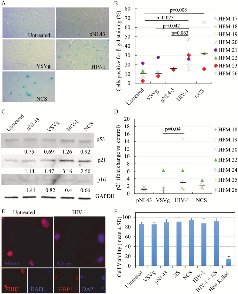
Figure 1.
Senescence biomarkers in microglia post-HIV-1 infection. SA-β-gal activity was detected in human fetal microglia (HFM) exposed to HIV-1 pseudotypes or controls. Representative pictures of microglia cultures (A) and summarized data from multiple cases (B), demonstrate increased SA-β-gal activity in microglia post-HIV-1 infection. Total of eight cases were used but not all experimental conditions could be done with all cases [group size: untreated, n = 8; pNL43, n = 4; VSVg, n = 5; VSVg pNL43, n = 7; Neocarzinostatin (NCS), n = 3]. Black bars represent the median for each experimental group. P values were calculated using the non-parametric Wilcoxon’s signed rank test (two-tailed). Representative Western blot for detection of p53, p21, and p16 levels, and GAPDH as loading control, in microglia (C). Densitometry values were normalized to the loading control and then expressed as fold-change with respect to untreated cells. Summarized Western blot data for p21 expression from multiple cases (D). Total of seven cases were used, but not all experimental conditions could be done with all cases (group size: untreated, n = 7; VSVg, n = 6; pNL43, n = 3; VSVg pNL43, n = 7; NCS, n = 4). Black bars represent the median for each experimental group. 53BP1 foci formation was evaluated post-HIV-1 infection (E). Immunofluorescence staining for DNA damage foci was performed, and 53BP1 (red) and nuclei marker DAPI (blue) are shown. Viability data of multiple microglia cases for each experimental group (F). The percentages of nucleated and viable cells were measured in singlicate (single measurement per condition per case) and are shown as mean ± SD. Heat-killed cells were used as positive control. Nucleosides (NS) were used at 250 nM. NCS concentrations were 1.0 µg/mL (HFM22-24) or 0.5 µg/mL (HFM25, 26).

