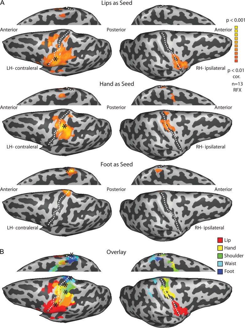
Figure 5.
Whole-brain functional connectivity analysis reveals highly symmetrical networks. (A) Statistical parametric maps of the random effect whole-brain functional connectivity analysis of the resting BOLD signal from the lips (upper), hand (middle) and foot (lower) seeds in the contralateral hemisphere (marked by asterisks) show that the peak connectivity in the ipsilateral hemisphere is localized in the homologous area of each contralateral seed. (B) Overlay of the functional connectivity maps of all 5 body segment seeds shows the reconstruction of the ipsilateral homunculus.

