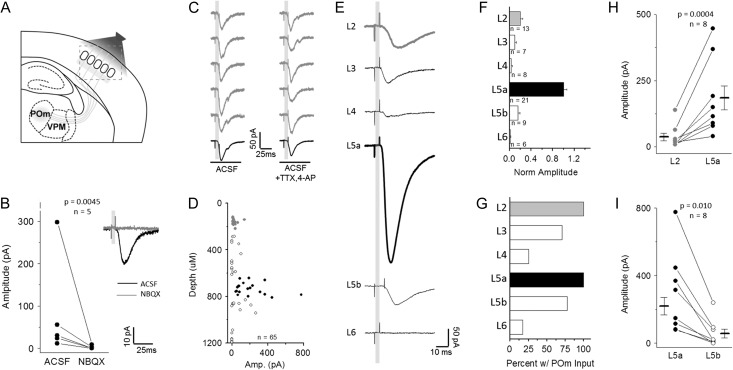
Figure 1.
Excitatory neurons in all cortical layers receive direct synaptic input from POm. (A) Schematic of experimental design. POm-targeted ChR2 viral injection in POm neurons labeled axons in barrel cortex. (B) EPSCs evoked by POm light stim in L2 Pyr neurons, eliminated by addition of AMPAR antagonist NBQX. About 5 consecutive trials (gray) followed by response average for 10 trials. (C) Short latency EPSCs in L2 Pyr neurons are direct from POm axons. EPSCs persist with addition of voltage-gated Na+ channel antagonist TTX and K-channel antagonist 4-AP. (D) Direct POm-evoked EPSCs in cortical Pyr neurons in TTX and 4-AP plotted by depth from pia surface (gray: L2, black: L5a). (E) Average cell response (10 trials) for a representative cell in each cortical layer, defined by cytoarchitecture and depth. (F) Across-layer comparison of amplitude normalized to the average L5a response in each slice to account for across-preparation variance in ChR2 expression. (G) Percent of recorded cells in each layer that receive direct POm-evoked EPSCs. (H) L5a Pyr neurons receive larger direct input than in L2. Connected points represent a comparison of the average of all responses recorded in L2 or L5a neurons from a given animal. Data points and n value are animal averages and a paired t-test is used. (I) L5a Pyr neurons receive larger direct POm input than L5b neurons; same as (H).

