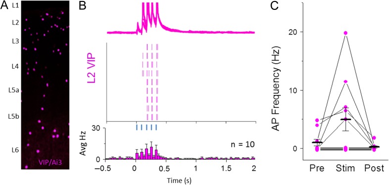
Figure 7.
A subset of L2 VIP GABAergic neurons contribute to POm-evoked spiking pattern of L2 5HT neurons. (A) Schematic of VIP-expressing GABAergic neurons throughout a cortical column in S1 imaged with YFP fluorescence. (B) Top: Overlay of 10 consecutive POm-evoke responses in mACSF (5 light pulses, 80 ms ISI, max intensity). Middle: Raster plot showing spike times for 10 consecutive traces shown for sample cell. Bottom: average peri-stimulus firing histogram for all L2 VIP neurons recorded. (C) Firing rate quantification for 500 ms bins before, during, and after POm stimulation for all VIP neurons recorded. Cells with no evoked spikes are slightly offset for visibility.

