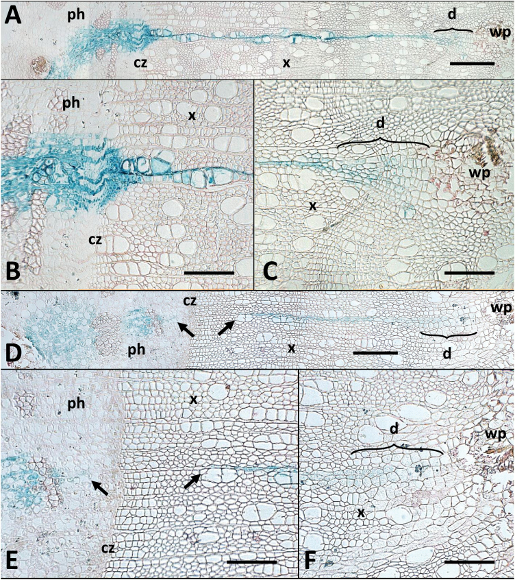
Fig. 2.
Micrographs of cross-sections showing histological detail of a continuous ‘full’ cambial sector and a split ‘lost initial’ sector. Micrographs show a continuous ‘full’ cambial sector (p46-3 in Supplementary Table S1; A–C) extending from the wound parenchyma (wp), through the xylem (x) and the cambial zone (cz) into the phloem (ph); and a split ‘lost initial’ sector (p33-11 in Supplementary Table S1; D–F) in which the phloem and xylem portions are radially aligned but separated across the cambial zone (cz). The gap across the cambial zone is indicated by arrows in (D) and (E). The dedifferentiation zone (d) is marked in (A), (C), (D), and (F). Scale bars: (A, D)=400 μm; (B, C, E, F)=200 μm.

