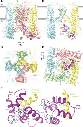
Fig. 1. Structure of the hTRPV6-CaM complex.

(A to D) Side (A and B), top (C), and bottom (D) views of hTRPV6-CaM with hTRPV6 subunits (A to D) colored cyan, green, yellow, and pink, and CaM colored purple. Calcium ions are shown as green spheres. In (B), only two of four hTRPV6 subunits are shown, with the front and back subunits removed for clarity. Side chains of TRPV6 residue W583 and CaM residue K115 are shown as sticks. (E) Expanded view of CaM bound to the proximal and distal portions of the TRPV6 C terminus. Side chains of CaM residues coordinating calcium ions in the EF hand motifs and K115 are shown as sticks.
