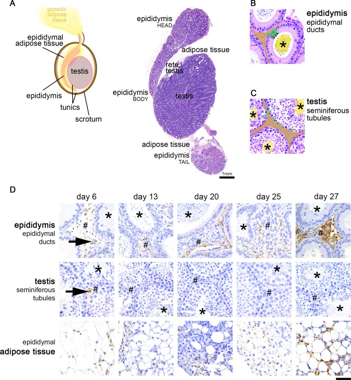
Fig 2. Histological features and parasite distribution in the male reproductive system.
A. Graphical representation and histological image of the external organs and tissues of the male reproductive system in the mouse (naïve, non-infected), corresponding to testis, the fibrous tunics that enclose the testis (tunica albuginea and tunica vaginalis), the epididymis, epididymal adipose tissue, and scrotum (skin) that confines all these organs/tissues. B. High magnification of the epididymis, which consists in ducts lined by a monolayer of epithelial cells (green). Tight-junctions in the apical side of these cells (black line) form the blood-epididymis barrier, which protects the numerous spermatozoa present in the lumen of epididymal ducts (asterisk, yellow). The stromal compartment (interstitium, orange), corresponding to connective tissue that contains blood vessels, is outside the blood-epididymis barrier. Immune cells (pink) also localize in the basal half of the intercellular space of the epithelia. C. Testis are composed by seminiferous tubules, lined by Sertoli cells (green). These cells are connected by tight-junctions (black line), forming the blood-testis barrier. The barrier protects spermatids and spermatozoa that accumulate in the lumen of the tubules (asterisk, yellow) from being recognized by the immune system. Outside the barrier is the stromal compartment (interstitium, orange), corresponding to connective tissue that contains blood vessels and Leydig cells. D. In the epididymis, parasites were detected inside the vessels (black arrow) but the great majority were extravascular, localized in the stroma (#) surrounding the epididymal ducts. No parasites were observed in the lumen of these ducts (asterisk). The testis had in general very few parasites. No parasites were found inside the seminiferous tubules (asterisk) nor in the stromal compartment (#) throughout the course of the infection, although at day 6 few trypanosomes were detected inside the vessels of the stroma of the testis (black arrow). The epididymal adipose tissue showed presence of parasites at all time-points post-infection. Hematoxylin and eosin of the male reproductive system of a naïve mouse (B, C). Anti-VSG immunohistochemistry of the testis, epididymis and epididymal adipose tissue of T. brucei-infected mice (n = 4 to 6 per time-point), DAB counterstained with hematoxylin (D), original magnification 40x (bar, 50μm).

