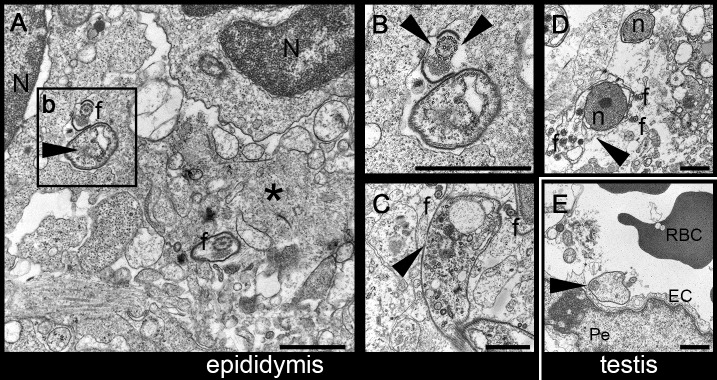
Fig 5. Morphological changes in T. brucei ultrastructure in late stages of the infection.
A. Transmission Electron Microscopy (TEM) of epididymis 27 days post-infection with T. brucei. Trypanosome cell bodies (black arrowhead) and flagella (f), admixed with cell debris, extracellular matrix proteins (asterisk) and associated with inflammatory mononuclear cells (N, nuclei). B. Detail of the trypanosome in Fig 5A showing a flagellum with an interrupted VSG coat (black arrowhead). C and D. Trypanosomes displaying severe signs of degeneration, with loss of VSG coat (black arrowhead), and admixed with numerous free flagella (f) and parasite nuclei (n). E. Trypanosome (black arrowhead) located inside the lumen of a vessel, in the testis (RBC, red blood cell; EC, endothelial cell; Pe, pericyte). Bar, 1μm. Mice, n = 2.

