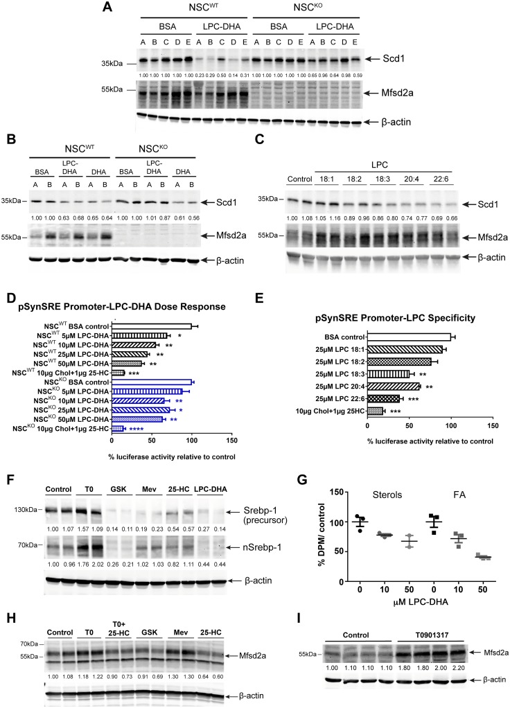
Fig 5. LPC-DHA inhibits Srebp activity and de novo lipogenesis and Mfsd2a is regulated by Srebp.
(A) Western blot analysis of Scd1 and Mfsd2a expression in NSCWT and NSCKO treated with or without 50μM LPC-DHA. Scd1 expression in NSCWT cells decreased with LPC-DHA treatment, but to a lesser extent in NSCKO cells. β-actin served as a loading control. Capital letters above each lane represent biological replicates for each genotype (n = 5 for NSCWT and NSCKO). (B) Western blot analysis of Scd1 and Mfsd2a expression in of NSCWT and NSCKO treated with or without 50 μM LPC-DHA or 50 μM DHA. DHA works in an Mfsd2a-independent manner to decrease Scd1 expression to a similar extent in NSCWT and NSCKO cells. β-actin served as a loading control. Capital letters above each lane represent biological replicates for each genotype (n = 2 for NSCWT and NSCKO). (C) Specificity of LPC species for down-regulation of Scd1. Western blot analysis of Scd1 and Mfsd2a expression in NSCWT treated with or without 50 μM each of LPC18:1, LPC18:2, LPC18:3, LPC20:4, or LPC22:6. Scd1 expression is more greatly reduced with increasing unsaturation of LPC-PUFAs. Mfsd2a expression remained relatively unchanged. β-actin served as a loading control. Technical replicates were carried out for each LPC treatment. (D) Dose-response effect of LPC-DHA on Srebp transcriptional activity. NSCWT and NSCKO cells were transfected with pSynSRE and treated with or without increasing concentrations of LPC-DHA, as indicated, or 25-hydroxycholesterol and cholesterol, as a positive control for repression of Srebp activity. The bar chart shows percent luciferase activity to respective untreated control as mean ± SE. LPC-DHA inhibited Srebp transcriptional activity in a concentration-dependent fashion in NSCWT but not NSCKO cells. Three technical replicates were carried out for each treatment condition. *p < 0.05; **p < 0.01, ***p < 0.001; ****p < 0.0001. (E) Specificity of LPC species for down-regulation of Srebp transcriptional activity. NSCWT cells transfected with pSynSRE and treated with or without 25 μM each of LPC18:1, LPC18:2, LPC18:3, LPC20:4, or LPC22:6 or 25-hydroxycholesterol and cholesterol (sterols). The bar chart shows percent luciferase activity to untreated controls as mean ± SE. Sterols inhibit Srebp and were used as a positive control for inhibition of Srebp transcriptional activity. Increased repression of Srebp transcriptional activity relative to control was observed with increasing unsaturation of LPC-PUFAs, consistent with effects observed for down-regulation of Scd1 expression shown in panel (C). The bar chart shows percent luciferase activity to untreated control as mean ± SE. Three technical replicates were carried out for each treatment condition. **p < 0.01, ***p < 0.001. (F) LPC-DHA reduces nuclear Srebp-1 levels. Western blot analysis of Srebp-1 expression in NSCWT treated with or without 50 μM of LPC-DHA. Activators and inhibitors of Srebp-1 processing and expression were used as controls to benchmark the effect of LPC-DHA. T0901317 (T0), an LXR agonist; GSK2033 (GSK), an LXR antagonist; Mevastatin plus mevalonic acid (Mev) inhibits HMG-CoAR activity; 25-hydroxycholesterol plus cholesterol (25-HC) inhibits Srebp-2 processing. β-actin served as a loading control. Technical replicates were carried out for each treatment condition. (G) LPC-DHA treatment reduced biosynthesis of sterols and fatty acids (FAs). [14C]-acetate lipogenesis assay was performed on NSCWT cells. NSCWT cells were treated with either 10 μM or 50 μM of LPC-DHA for 16 hours followed by supplementing cell media with [14C]-acetate for 2 hours. Sterols and fatty acids were purified as described in Materials and methods and quantified by scintillation counting. Uptake is expressed as mean percent DPM of untreated cells ± SE. Sterol synthesis was significantly reduced in 10 μM LPC-DHA–treated NSCWT relative to control (*p < 0.05). Fatty acid synthesis was significantly reduced in 50 μM LPC-DHA–treated NSCWT relative to control (**p < 0.01). Two to three technical replicates were carried out for each treatment condition. (H) Mfsd2a levels are regulated by Srebp activity. Western blot of Mfsd2a expression in NSCWT cells treated with or without activators or inhibitors of Srebp-1 and Srebp-2, as described in (F) above. β-actin served as a loading control. Technical replicates were carried out for each treatment condition. (I) LXR agonist treatment of mice enhances brain levels of Mfsd2a. Western blot analysis of Mfsd2a expression in of brain lysates of WT mice injected with 30 mg/kg/day T0901317 or vehicle control for 4 days. β-actin served as a loading control. Control, n = 4; T0901317, n = 4; biological replicates. Experimental data depicted in this figure can be found in S1 Data.

