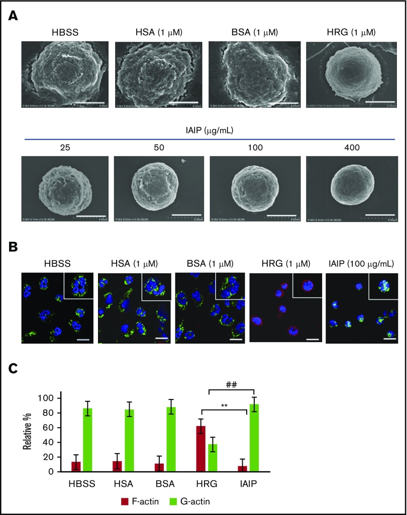
Figure 2.
Effects of IAIPs on surface structure and cytoskeletal arrangement in neutrophils. The purified human neutrophils were incubated with HBSS, HSA (1 μM), BSA (1 μM), HRG (1 μM), or different concentrations of IAIPs (25-400 μg/mL) in a 96-well microtiter plate for 1 hour at 37°C. (A) After fixation as described in “Methods,” scanning electron microscopic pictures of neutrophils were obtained. Typical cell appearance is shown from each group. All images were acquired at 12 000× magnification. Scale bars, 4.0 μm. (B) After 1-hour incubation at 37°C, the neutrophils were fixed with 4% PFA, and monomer actin (green) and F-actin (red) were stained immunohistochemically. All images acquired at 40× original magnification by confocal laser microscope. Typical cells are shown from each group. Insets represent the higher magnification picture of a single cell from each group. Scale bars, 10 μm. (C) The numbers of F-actin and G-actin dominant cells in panel B were counted in each group and were expressed as relative percentage. The results shown are the means ± SEM of 3 experiments. **P < .001, HRG F-actin vs IAIP F-actin; ##P < .001, HRG G-actin vs IAIP G-actin.

