
Figure 5: In vivo studies of localized chemo- radiotherapy.
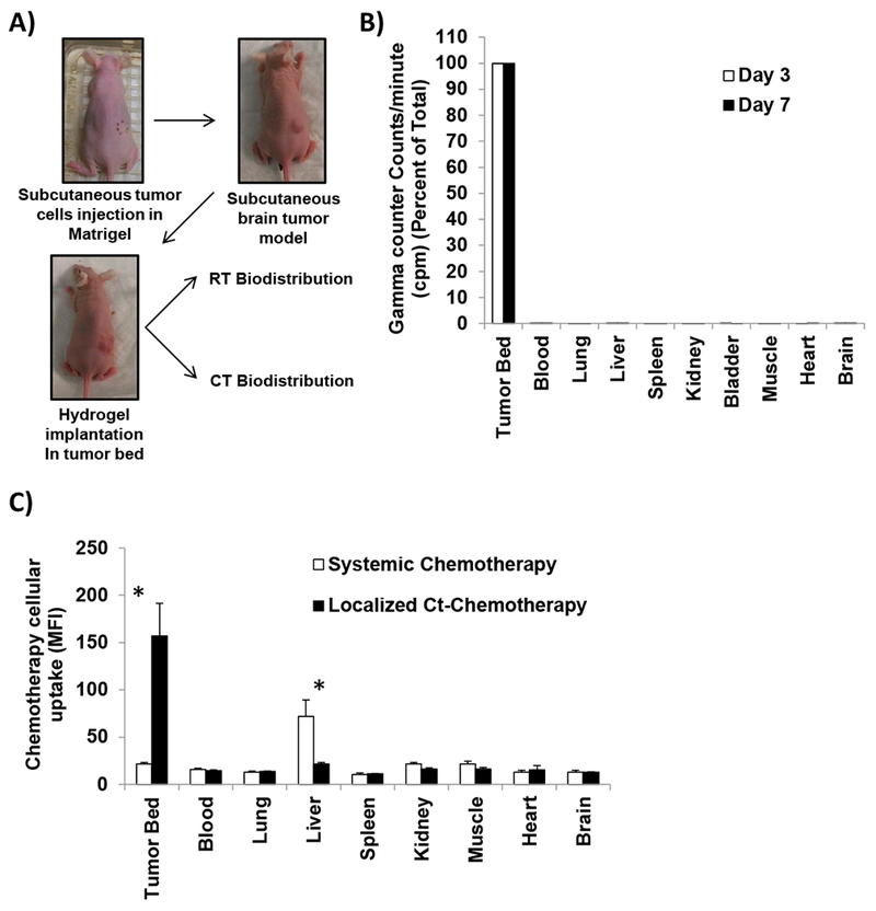
A) Design of in vivo subcutaneous brain tumor model, in which D54 GBM cells are injected in combination with matrigel, once tumors were palpable, mice were stratified into treatment groups where Ct hydrogels were implanted on top of the tumors and mice were followed by BLI twice a week, analyzed for radiotherapy and chemotherapy release. B) Radiotherapy bio-distribution in different organs after 3 and 7 days of implantation of radio-hydrogels measured by gamma counter (counts per minute, cpm) (n=3). C) Chemotherapy cellular uptake in different organs after 18 hours of implantation of localized chemo-hydrogels loaded with doxorubicin or i.v. systemic injection of same amount of doxorubicin (5mg/kg) measured by MFI in flow cytometry (n=3).