
Figure 2.
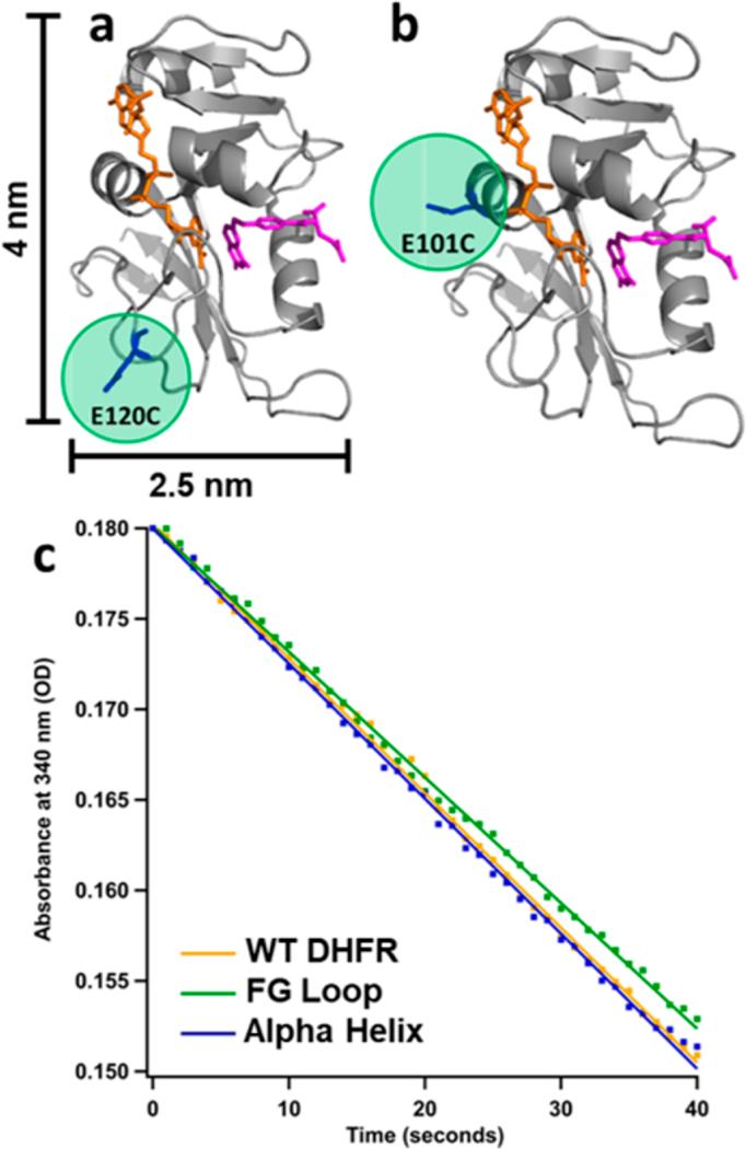
Location of cysteine mutation (highlighted in green) for (a) FG Loop mutant and (b) Alpha Helix mutant DHFR. Crystal structure from PDB 1RX2 with backbone shown in cartoon view (gray), substrate dihydrofolate in stick view (pink), and cofactor NADPH in stick view (orange). (c) Initial rate data for free protein when [DHFR] = 10 nM, [DHF] = 50 μM, and [NADPH] = 50 μM at 37°C. Yellow = WT DHFR. Green = FG Loop mutant. Blue = Alpha Helix mutant.