
Fig. 3.
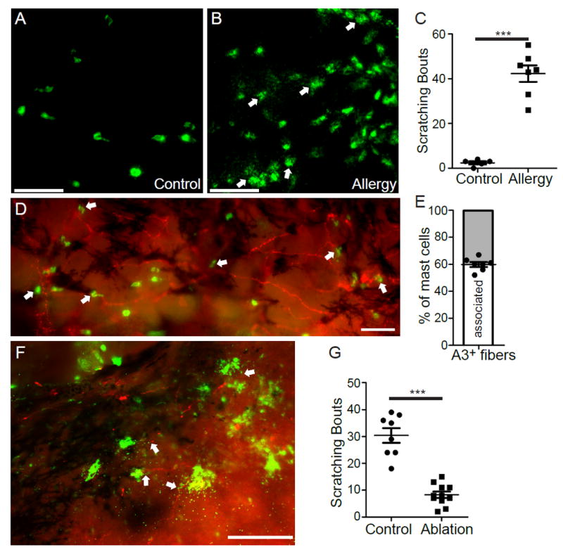
MrgprA3+ sensory neurons are required for mast cell-dependent allergic ocular itch. (A-B) Representative images of mast cells (stained with FITC-avidin, green) in the whole-mount conjunctivae of non-immunized (A) and immunized mice (B). Arrows indicate the granules released from mast cell bodies upon challenging with allergen ovalbumin. (C) Ocular scratching responses evoked by topical conjunctival application of allergen ovalbumin in non-allergic control mice (n=5) and allergic mice (n=7). Statistical analysis by two tailed Student’s t-test (***P=0.00003). (D) Representative image of the whole-mount conjunctiva from Mrgpra3tdTomato mice. Arrows indicated avidin-stained mast cells. (E) The proportion of mast cells closely associated with MrgprA3+ sensory fibers (n=7 conjunctival explants) (F) Representative image showing the interaction between released granules (green) and MrgprA3+ sensory fibers (red) in the conjunctiva upon allergen challenge, as indicated by arrows. All images shown are representatives of three independent experiments using tissues from at least 3 different mice. Scale bars: 50 μm. (G) Ocular scratching responses induced by mast cell-dependent ocular allergy in MrgprA3+ neuron-ablated mice (n=11) and control Rosa26HBEGF/+ mice (n=8). Statistical analysis by two tailed Student’s t-test (***P=0.0000003). All data are expressed as mean±s.e.m.