
Fig. 4.
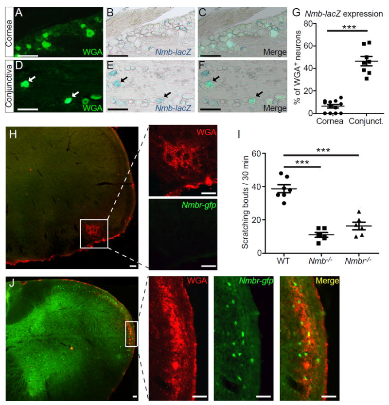
Central NMB/NMBR signaling is required for conjunctival itch transmission. (A-G) Representative images showing x-gal staining of Nmb-lacZ reporter in sections containing sensory neurons retrogradely labeled by Wheat Germ Agglutinin conjugated with Alexa Fluor™ 488 (WGA-488) from the conjunctiva or cornea ofNmbtm1.1(KOMP)Vlcg mice. Each dot in (G) represents one section image of trigeminal ganglia (n=6 TGs from three mice per group). Statistical analysis by two tailed Student’s t-test (***P=0.000008). (H, J) Representative images showing the absence or presence of Nmbr-GFP in the central projection area of corneal (H) or conjunctival afferent neurons (J). Scale bars: 50 μm. (I) Ocular scratching responses evoked by ocular allergy in Nmb-/- mice (n=5), Nmbr-/- mice (n=6) and WT (control) mice (n=7). Statistical analysis by one-way ANOVA followed by two tailed Student’s t-test (Nmb-/- vs. WT, ***P=0.000002; Nmbr-/- vs. WT, ***P=0.00004). All data are expressed as mean±s.e.m.