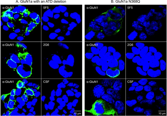
Figure 4.

GluN1 structural changes known to impair antigen binding by ANRE patient CSF IgGs also inhibit 5F5 and 2G6 binding. HEK293T cells expressing mutant GluN1 proteins were stained with a commercial anti‐GluN1 antibody (green), followed by 5F5, 2G6, or CSF (red). Nuclei were stained with DAPI. (A) The GluN1 amino terminal deletion mutant protein (ATD). (B) GluN1 with the N368Q mutation. Neither mutant GluN1 protein was recognized by 5F5, 2C6, or CSF. Scale bars = 10 μm.
