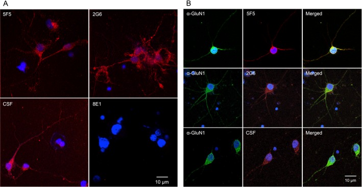
Figure 8.

The 5F5 and 2G6 mAbs bind GluN1 on rat hippocampal neurons. (A) Neurons were cultured for 14 days and stained with ANRE CSF or mAbs (red). Top left, 5F5. Top right, 2G6. Bottom left, CSF. Bottom right, 8E1. Nuclei were stained with DAPI. Scale bar = 10 μm. (B) Neurons were stained with ANRE CSF or mAbs (red), and costained with murine anti‐GluN1 antibody (green). Rows: Top, 5F5. Middle, 2G6. Bottom, CSF. Columns: Left, GluN1. Middle, CSF or mAbs. Right, Merged images. Nuclei were stained with DAPI. Scale bar = 10 μm.
