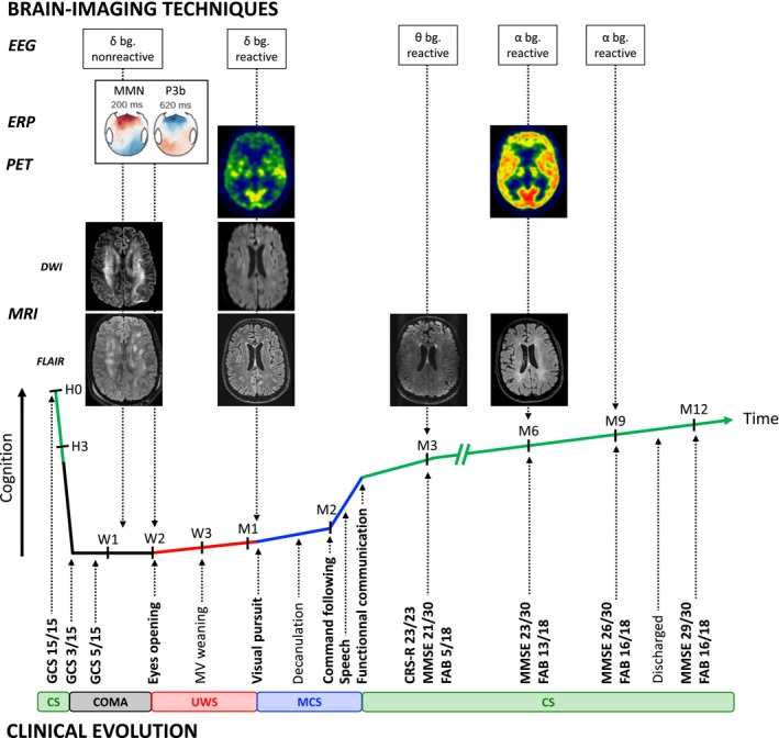
Figure 3.

Multimodal clinical and brain‐imaging dynamic of recovery. Legend. (Bottom) Longitudinal clinical follow‐up of the patient's recovery according to different scores assessing vigilance (Glasgow Coma Scale), consciousness (Coma Recovery Scale‐ Revised) and cognitive function together with pivotal behavior. (Top) Concurrent brain function according to different functional and structural brain imaging techniques (electroencephalogram, auditory event‐related potential, 18‐FDG PET‐TDM and MRI). bg, background rhythm; CRS‐R, Coma recovery scale ‐revised; CS, Conscious state; D, Day; DWI, Diffusion‐weighted imaging; EEG, Electroencephalogram; ERP, Event‐related potential; FAB, Frontal assessment battery; FLAIR, Fluid‐attenuation inversion recovery; GCS, Glasgow coma scale; H, Hour; M, Month; MCS, Minimally conscious state; MMN, Mismatch negativity; MMSE, Mini‐mental state examination; MRI, Magnetic resonance imaging; MV, Mechanical ventilation; PET, Positron‐emission tomography; UWS, Unresponsive wakefulness syndrome; W, Week.
