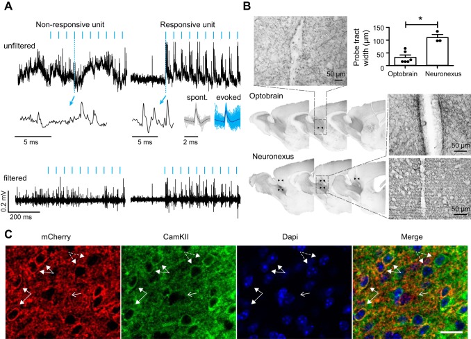
Fig. 2.
Absence of a light artifact and limited tissue damage. A: no photo-induced light artifact was observed in vivo. The anterior olfactory nucleus was transduced with rAAV2/7-CamKII-ChR2(L132C/T159C)-mCherry, and electrophysiological recordings were performed in awake head-restrained mice. Top and bottom graphs represent raw and filtered (600–6,000 Hz) signals, respectively, of a nonresponsive (left) and a responsive unit (right) recorded on the same tetrode during 20-Hz stimulation with 5-ms light pulses of blue light (470 nm). Blue arrows indicate a magnified view of a raw signal fragment during optical stimulation. Spontaneous (black) and light-evoked (blue) waveforms are represented for the responsive unit (cross-correlation ~98%). B: reduced insertional damage compared with that for a commercially available probe (NeuroNexus) with 125-µm optical fiber. Sagittal sections of diaminobenzidine staining for mCherry are shown surrounding the probe tract of both probes (indicated with black arrowheads). Insets show an enlargement of the probe tract of both devices. At top right, the mean width (±SD) of the probe tract is represented for the optoelectrode (n = 6) and NeuroNexus probe (n = 3). *P < 0.01 (one-way ANOVA, P < 0.05, df = 2). C: selective expression of ChR2-mCherry in CamKII+ cells. Confocal images show mCherry and CamKII expression at ×40 magnification. Example cells positive for both mCherry and CamKII are highlighted by closed arrows. Example cells positive for CamKII but not mCherry are highlighted by dashed arrows. Arrow with open head points to a cell that is negative for mCherry and CamKII. Scale bar, 30 µm.

